Menu ×

Book Appointment
Pay Online

Cat Scans

In our last column we covered ultrasound scanning as a way to image your pet. This week, we’re going to look at another way of seeing inside the animal body: the CAT Scan.  “CAT” is an abbreviation of “computed tomography”. This is derived from the Greek words “tome”, meaning “slice”, and “graphi” which means “to write”.  The reason this name is used is because CAT scans make images of the body in slices.  These slices are then assembled to make a study of the body.

A CAT scanner is a type of x-ray machine. It is very large and looks like a doughnut. Inside the doughnut is an x-ray tube and a semi-circle of sensors. The patient lies on a tray that can move in and out of the doughnut. Images are generated when X-rays pass through the body to the sensors in a rotating arc. As the tray moves in and out of the doughnut, these images are generated in slices. These slices are then transmitted to a computer to form a complete picture of the area under examination. The images can be two-dimensional or three-dimensional depending on the computer and the requirements of the vet.

The use of CAT scans has exploded in recent decades as the price of these machines has come down. It is now reasonably routine for pets to be scanned for a range of conditions. These conditions include fractures, spinal problems, tumours and virtually any other issue you can think of in the body. When we have an animal that is undergoing a CAT scan they generally require sedation or even full anaesthesia. The reason for this is that we cannot have an animal standing up and walking off a scanner in the middle of a scan! This means that when your pet undergoes a CAT scan they usually have to be admitted to hospital for at least a day.

CAT scans have some advantages over traditional X-ray images. CAT scans have better resolution than traditional radiographs and because a number of images are taken CAT scans avoid the superimposition of structures that can happen on a plain one-dimensional X-ray. This means that interpretation of these images can often give more information than would otherwise be achievable with a normal x-ray.

CAT scans also have some disadvantages. One disadvantage is the cost of the machine.  Purchase and operation of CAT scanners is expensive. Another disadvantage is the radiation dose that the patients can receive during the scan. Clinicians are aware of the need to reduce radiation exposure in patients. Therefore only patients which require a scan will undergo one.  The clinician must undertake a cost-benefit analysis before ordering a scan for any particular patient to balance the risk of the scan against the benefits of increasing the knowledge of a patient’s condition.

CAT scans have undoubtedly moved veterinary medicine forward over the past two decades. They have enabled us to see deeper into the body of an animal than we have ever been able to see before. They provide more information on cases that enable us to tailor treatments better to the individual animal and improve patient outcomes.  We hope that in the future costs will come down even further to broaden their use in general practice.

Cat Scan Or Lab Test Anyone?

There is a fairly old joke about a bloke going to a doctor complaining of a mystery illness.  The doctor is stumped so he turns to a cat who paws at the man, slowly shakes her head and walks off.  The doctor then asks a Labrador to give the man the once over.  The Labrador sniffs the patient, shakes his head, and leaves the room.  The doc then hands the patient a bill for £1000.  The bloke is furious – he exclaims – you didn’t do anything except let a few animals into the consultation room.  The doctor calmly explains that he did.  He requested a cat scan and a lab test!

OK, so perhaps the joke isn’t as funny as I thought when I first heard it.  But this week in the news we saw a great example of how technology is changing the world of medicine.  Headlines related how Google Health had used a computer to diagnose particular cancers as successfully as experienced and skilled doctors.  It is an encouraging example of combining powerful technology and medical skills to improve patient outcomes.  This got me thinking – could we use tech to help our animal patients?

Change is coming.  And it’s exciting.  One obvious area that we can use computing power to improve diagnosis is in assessing x-rays, scans and test results.  When vets look at these tests, they are evaluating them for patterns.  Certain patterns mean that their patients are suffering from this or that disease.  If we can train a computer to look for the same patterns, then we can increase the accuracy and rate of diagnosis.  A computer is very good at repeating the same task over and over.  The computer will not replace the vet, but will add and extra opinion that can be very useful in tricky cases.

Another area which is promising is patient-side diagnostics for disease.  If we could take a sample from a patient and run a test telling us what bug is causing that disease within 5 minutes, then we can get treatment started sooner.  E.g. at the moment it takes us about a  week to culture bacteria, and find out what antibiotic kills them.  If we can speed this up, then we can treat the disease quickly, and also save antibiotics by only using the correct one for the first time, every time.

3D printing is perhaps another technology that is already changing how things are done in surgery.  Orthopaedics is the science of fixing bones.  In order to fix broken bones we often have to fix them together with bits of metal – plates and pins.  Every patient is different, so it can be hard to find the correct size of pin to suit the patient.  Now, though, we can scan the patient’s bones, measure their length and actually print an implant that is tailor-made for the patient.  This can improve the chances of success of any surgery.

These are just a few areas that technology is likely to change patient care in veterinary medicine.  It will be interesting to revisit this article in a few years to see if any of my predictions have come true.  Watch this space!

Calf Pheumonia – Autumn 2024

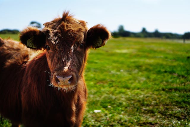
It’s that time of year again for the farmers of Lecale – the time to start preparing your herds for housing during the winter months. As covered in previous issues, a big threat to the health of cattle at housing is calf pneumonia. We have seen a lot of pneumonia in the past weeks even in cattle grasinging in the fields. We suspect that the culprit in this case is lungworm, and we will cover that disease in another article. For this article I would like to discuss the spread of viral and bacterial pneumonia as a result of close contact of animals at housing.

Calf pneumonia, also known as bovine respiratory disease (BRD), is a prevalent and potentially devastating condition that can affect young cattle, especially in their first few months of life. This illness not only impacts the health and growth of the calves but can also lead to significant economic losses. At Downe Vet Clinic, we advocate for a proactive, three-pronged approach to prevent calf pneumonia: maintaining excellent hygiene, providing balanced nutrition, and implementing a strategic vaccination program.

Understanding Calf Pneumonia
Calf pneumonia is caused by a combination of viral and bacterial pathogens that exploit stressors such as poor ventilation, sudden changes in weather, or inadequate nutrition. These factors weaken a calf’s immune system, making it more susceptible to respiratory infections. Early and consistent preventive measures are crucial in reducing the incidence and severity of this disease.

The Importance of Hygiene: Good hygiene is fundamental in preventing calf pneumonia. By maintaining a clean environment, you reduce the number of pathogens that calves are exposed to, thereby lowering the risk of infection.

Clean and Dry Housing: Ensure that calf pens are regularly cleaned, dry, and well-bedded. Wet, dirty bedding can harbour harmful bacteria and viruses that lead to respiratory problems. Regularly removing soiled bedding and replacing it with fresh material is essential.

Proper Ventilation: Adequate ventilation is critical to minimising the buildup of ammonia and moisture in calf housing, both of which can irritate the respiratory tract and increase vulnerability to infections. Ensure good airflow without creating drafts that could chill young calves.

Routine Disinfection: Regularly disinfect feeding equipment, water troughs, and housing areas to reduce the pathogen load in the environment. Use effective disinfectants and adhere to recommended protocols to ensure thorough cleaning.

Colostrum Management: Providing high-quality colostrum within the first few hours of life is vital for boosting calves’ immunity. Proper handling and feeding techniques are necessary to prevent contamination and ensure that calves receive the full benefits of colostrum.

Blue Green Algae Is Back

The dog walkers among you may have noticed signs in local beauty spots that Blue Green Algae is back in our lakes and ponds. But what is this stuff, and is it dangerous for your dog?

Blue Green Algae is not actually algae at all. It is a type of bacteria called cyanobacteria. It is called “blue / green algae because it looks like, well, Algae that is greenish blue. “Cyano” means blue in Greek, and the cyanobacteria are usually blue or green when seen in large numbers by the human eye. Cyanobacteria live in water, like lakes and ponds. Usually they pose no problems as their numbers are small. However, under certain conditions and during certain times of the year, cyanobacteria may produce an “Algal Bloom”. You may notice this as a green / blue or even mud-red colour on the water of your local lake or pond. These algal blooms can cause problems to fish, animals and even people.

The problem with algal blooms is that the bacteria can produce toxins (known as cyanotoxins). These toxins can have a wide range of effects on animals, people and fish. Symptoms vary from rashes to causing vomiting, diarrhoea and even organ failure in large doses. The symptoms an animal can develop depends on lots of factors: how long was the animal in contact with the water, did it drink the water, what type of cyanobacteria are present? So it can be hard to predict if an animal will be sick and what symptoms they will have after coming into contact with cyanotoxins. The best advice is to avoid contact with any water where an algal bloom is suspected. Local authorities are vigilant about posting notices near affected lakes and ponds.

If your dog loves swimming and unwittingly jumps into water where there is an algal bloom, there are a few steps you can take. Firstly call your dog out of the water. It is prudent to take sensible precautions like wearing gloves / goggles and refraining from entering the water yourself. Wash the coat of the dog with fresh, clean water to stop the dog licking his coat. Call your vet. They can make your dog vomit just in case the dog has ingested any water. The sooner the better. In more serious cases your vet may admit your pet for blood tests and supportive treatment.

In most of the cases we have treated the outcome is good. However, prevention is better than cure, and adhering to local restrictions is the best advice. Blue Green algae are normally an important part of our ecosystem, but algal blooms are not. Enjoy the summer with your dog, and stay safe.

Join our newsletter today

Sign up to get the lastest news & offers

Loading