The dog walkers among you may have noticed signs in local beauty spots that Blue Green Algae is back in our lakes and ponds. But what is this stuff, and is it dangerous for your dog?
Blue Green Algae is not actually algae at all. It is a type of bacteria called cyanobacteria. It is called “blue / green algae because it looks like, well, Algae that is greenish blue. “Cyano” means blue in Greek, and the cyanobacteria are usually blue or green when seen in large numbers by the human eye. Cyanobacteria live in water, like lakes and ponds. Usually they pose no problems as their numbers are small. However, under certain conditions and during certain times of the year, cyanobacteria may produce an “Algal Bloom”. You may notice this as a green / blue or even mud-red colour on the water of your local lake or pond. These algal blooms can cause problems to fish, animals and even people.
The problem with algal blooms is that the bacteria can produce toxins (known as cyanotoxins). These toxins can have a wide range of effects on animals, people and fish. Symptoms vary from rashes to causing vomiting, diarrhoea and even organ failure in large doses. The symptoms an animal can develop depends on lots of factors: how long was the animal in contact with the water, did it drink the water, what type of cyanobacteria are present? So it can be hard to predict if an animal will be sick and what symptoms they will have after coming into contact with cyanotoxins. The best advice is to avoid contact with any water where an algal bloom is suspected. Local authorities are vigilant about posting notices near affected lakes and ponds.
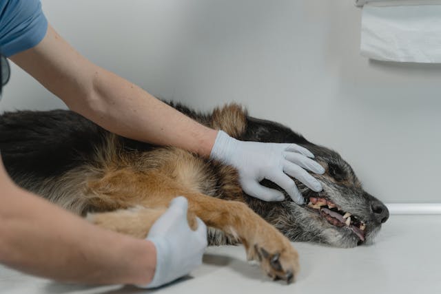
If your dog loves swimming and unwittingly jumps into water where there is an algal bloom, there are a few steps you can take. Firstly call your dog out of the water. It is prudent to take sensible precautions like wearing gloves / goggles and refraining from entering the water yourself. Wash the coat of the dog with fresh, clean water to stop the dog licking his coat. Call your vet. They can make your dog vomit just in case the dog has ingested any water. The sooner the better. In more serious cases your vet may admit your pet for blood tests and supportive treatment.
In most of the cases we have treated the outcome is good. However, prevention is better than cure, and adhering to local restrictions is the best advice. Blue Green algae are normally an important part of our ecosystem, but algal blooms are not. Enjoy the summer with your dog, and stay safe.

